


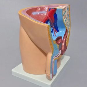
Female Pelvis Anatomy (Median Section)
Female Pelvis Anatomy (Median Section)
Introduction to Female Pelvis Anatomy Chart
Anatomy charts serve as essential tools in medical education, providing visual representations of complex structures within the human body. This article focuses on the female pelvis anatomy chart, specifically its median section diagram, which offers valuable insights into the intricate relationships between various anatomical structures.
An Overview of the Female Pelvis Anatomy
The female pelvis is a complex region consisting of bones, muscles, ligaments, and internal organs. It serves multiple functions, including supporting the weight of the upper body, facilitating childbirth, and protecting vital organs. Understanding the anatomy of the female pelvis is crucial for healthcare professionals, researchers, and students alike.
Understanding the Importance of Medical Education Tools
Medical education tools like anatomy charts play a vital role in teaching and learning about the human body. They help students visualize and comprehend complex anatomical structures, making it easier to grasp their functions and relationships. In this article, we will explore how the female pelvis anatomy chart can be used as an effective educational tool.
Exploring the Median Section Diagram
The median section diagram of the female pelvis provides a comprehensive view of the pelvic structures from a midline perspective. This section includes the bones, muscles, ligaments, and internal organs, allowing for a better understanding of their spatial relationships.
Identifying Key Structures in the Female Pelvis
Several key structures are visible in the median section diagram of the female pelvis, including:
- Pelvic bones (iliac crest, pubic symphysis, sacrum)
- Muscles (levator ani, obturator internus, piriformis)
- Ligaments (round ligament, cardinal ligament, uterosacral ligament)
- Internal organs (uterus, vagina, bladder, rectum)
Understanding the Relationship Between Structures
The median section diagram helps illustrate the spatial relationships between these structures, allowing students to better understand their functions and interactions. For example, the uterus sits between the bladder and rectum, while the levator ani muscles support the pelvic floor.
Using the Female Pelvis Anatomy Chart as a Medical Education Tool
The female pelvis anatomy chart serves as an effective educational tool for teaching students about the female pelvis anatomy. By incorporating this chart into teaching methods, educators can enhance learning outcomes and help students develop a deeper understanding of the subject matter.
Incorporating the Chart into Teaching Methods
Instructors can use the female pelvis anatomy chart in various ways, such as:
- Lectures: Presenting the chart during lectures to illustrate key concepts and structures
- Hands-on activities: Using anatomical models or cadavers to reinforce the information presented in the chart
- Group discussions: Encouraging students to analyze and discuss the relationships between structures
Enhancing Learning Outcomes with the Female Pelvis Anatomy Chart
By integrating the female pelvis anatomy chart into their teaching methods, instructors can help students achieve better learning outcomes, such as:
- Improved comprehension of complex anatomical structures
- Enhanced ability to visualize spatial relationships between structures
- Increased confidence in applying knowledge to real-world scenarios
In conclusion, the female pelvis anatomy chart, specifically its median section diagram, serves as an invaluable tool for medical education. By understanding the importance of these charts and incorporating them into teaching methods, educators can effectively enhance learning outcomes and foster a deeper understanding of female pelvis anatomy among students.

LABOTECH TRADING
Review

You May Also Like

Creative New Hide Private Money Tibetan Medicine Storage Box With Cable Fake Charging Treasure Mobile Power Organizer Case Gifts

Portable Medicine Box Health Supplement Organizer Sealed Pill Container Travel-friendly Medicine Storage by Sanrio Kawaii
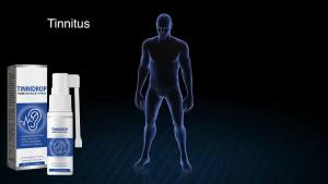
OTBK Tinnidrop: Earwax Cleaning & Tinnitus Relief Spray

💯Antartic Krill Oil Gel Candy 60 Capsules Astaxanthin Supplements Omega-3 Astaxanthin Supplement pill krill for Cardiovascular Phosphorus-Rich Nutritional Supplement

Crystal C Alkaline Soduim Ascorbate non Acidic 2 boxes Trial Package

Blood Glucose Control Cream & Diabetes Care: A Comprehensive Guide

Black Wheelchair Side Pouch Portable Armrest Pouch 3 Pockets Wheelchair Side Bag For Wheelchair

Best Pro Rechargeable Digital Hearing Amplifier In The Canal Hearing Aids Mini CIC Hearing Aid With Noise Canceling
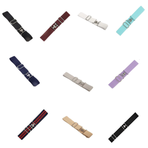
High Elasticity Equestrian Belt Adjustable Length Shark Buckle Waist Strap For Horseback Riding And Fashion Enthusiasts

Original Psoriasis Cream Skin Care Cream Psoriasis Skin Cream Dermatitis Eczematoid Eczema Ointment Treatment 20g Psoriasis Eczema Itchiness Organic Skin Ointment Natural Herbal Multi Plant Extract Anti Bacterial Anti Fungal 👍
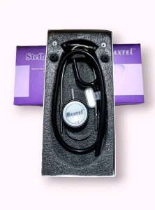
Baxtel Deluxe Stethoscope: A Durable & High-Quality Medical Tool